

Physical Therapy for the Middle Back
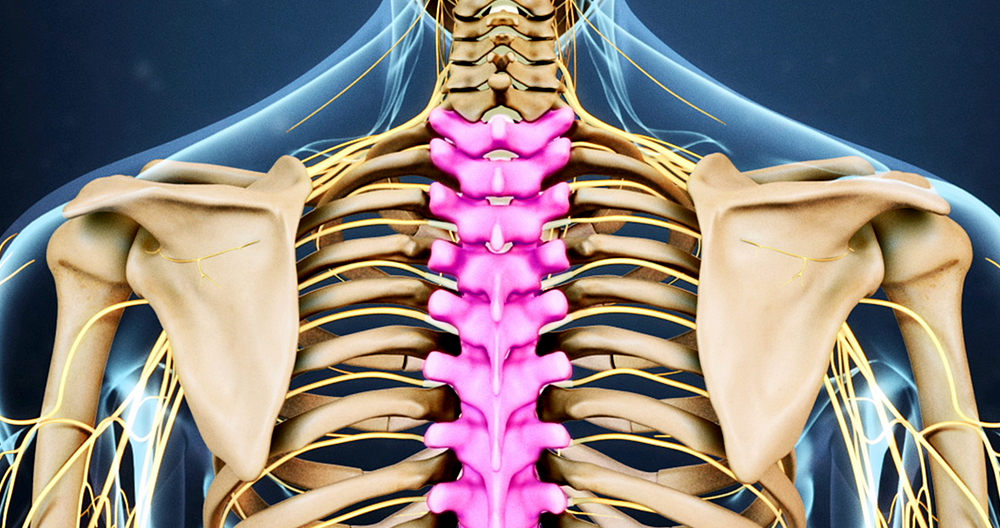
Why do I have this problem? Most thoracic disk herniations result from wear and tear. This is called “degeneration.” As a disc’s nucleus gets older, it cracks and tears. These injuries can often be repaired with scar tissue. As the annulus becomes weaker over time, the nucleus could herniate from the damaged annulus. T11, T12 are areas that are most susceptible to spinal degeneration. T12 is the area where the lumbar, thoracic and cervical spines meet. This link is easily damaged by daily activities like twisting and bending. Here is where most thoracic disk herniations occur.
A thoracic Disc can sometimes herniate suddenly (an acute injury). Thoracic discs can rupture due to an accident, fall, or other circumstances. A herniated disc in the thoracic area can result from a sudden twisting of the middle back.
Thoracic spinal nerve diseases can lead to thoracic disk herniation. Scheuermann’s patients may have thoracic disc herniations. Even though evidence isn’t conclusive in this instance, it appears that Scheuermann’s disease patients could have multiple herniated discs.
A herniated thoracic disc can cause damage to the spinal cord. The narrowest portion of the spinal canal is in the thoracic vertebrae. This can be very dangerous. Anything that takes up space within this canal can cause injury. Most disc herniations within the thoracic spinal canal push to one side, rather than deflecting. The disc material is pushed straight to the spinal cord. A herniated disc can cut off blood supply to the spinal chord. The critical zone, T4-9 of the Thoracic Spine, can be affected by discs that protrude in this area. This could result in a cut to the spinal cord’s blood supply. This can cause paralysis and weakness in your legs.
SYMPTOMS
What does this condition look like? Different symptoms can be seen in a case of thoracic disc herniation. The symptoms will vary depending on the severity of the injury, the size and location of the disc, as well as the pressing and pressure levels.
Pain is the most common symptom. While the pain is typically located around the injured disk, it can also spread to the side or both of your mid-back. A common sensation is a pain in the chest. Some patients may feel pins, pins, and numbness. Others may feel weak in their arms and legs. A disc material pressing against the spine can cause problems in the bowel and bladder.
Disc herniations may also cause pain in other areas than the spine. The pain caused by herniations at the upper thoracic and cervical spines can radiate to either one or both of your arms. A herniation in the middle of the thoracic spinal vertebrae can cause pain that radiates to the chest or abdomen. A lower thoracic disk herniation can cause pain in your groin, lower legs, and mimic kidney problems.
DIAGNOSIS
How can doctors diagnose the problem? For diagnosis, it is necessary to take a full history and perform a physical examination. Your symptoms will be examined by your doctor. They will also ask you about how the problem affects your daily activities. Your doctor will ask you questions like where do you feel pain, and if you feel numbness in the legs or your legs. Your doctor will ask you about your activities or other positions that may be causing your symptoms. Next, the doctor will examine your back to see if you have back pain or if you have back movements that are causing it. Also, your skin sensation, muscle power and reflexes will be assessed.
X-rays show the bones. Unless one of the discs is calcified or multiple discs are present, X-rays will not show the discs. This is essential in diagnosing thoracic Disc Herniation. If the calcified disc is visible in your spinal canal, this can be used to confirm disc herniation. It is unknown why a thoracic disk sometimes hardens from calcification. However, it could be due to an injury in the past.
Magnetic resonance imaging (MRI), the best way to diagnose a herniated thoracic back, is magnetic resonance imaging. Magnetic resonance imaging uses magnetic waves (MRI) to visualize the soft tissue. It allows you to see the discs clearly and determine if any have herniated. The machine captures images that look like slices of the area in which your doctor is concerned. This test does NOT require dye or needles. Many people may have thoracic back pain, even though they don’t experience symptoms. Doctors suggested that thoracic back herniations may not be associated with symptoms.
Myelography was used by doctors to diagnose thoracic Disc Herniations before MRI. Myelography is only half the way to diagnose this condition. Myelography uses X-rays. The spinal canal is treated with a special dye. The dye can be seen on an X Ray. This dye allows a doctor to see if the disc is pressing into a spinal canal.
A computed tomography scan (CT scanning), may be requested. This is a detailed X Ray, which allows doctors to view the tissues in images that look almost like slices. Images give additional information about calcified disks. Doctors might combine the CT scan and myelography. Myelography dye is used during a CT scan to highlight the spine or nerves. The myelography color can be used to improve diagnostics and accuracy in a CT scan. It is used to diagnose a herniated, bulging or bulging thoracic disk.
Doctors use MRI to diagnose thoracic disc herniations. Doctors may also use CT scans, myelography, or MRI to diagnose herniated cervical discs.
TREATMENT
What are your options for treatment?
Nonsurgical Treatment
Even if the herniation is not large, doctors will closely monitor patients suffering from symptoms of a herniated cervical disc. If the disc is placing pressure on the spine chord or blood vessels, severe neurological symptoms could develop quickly. These situations call for immediate surgery. Unless your condition becomes severe or rapidly worsening it is best to seek nonsurgical treatment.
In the beginning, your doctor may recommend immobilizing it. You can calm inflammation and reduce pain by keeping the back still for a few minutes. Sometimes, it may be necessary to rest for a few days. This is because lying on your back could cause pressure to nerves or discs. Doctors advise against bed rest. They encourage patients to engage in normal activities. Patients can also use pain to determine how much is too much. Braces can also be used to immobilize your back for up one week.
Some medications may be prescribed to patients suffering from thoracic or cervical disc herniation by their doctors. For patients suffering from thoracic disc herniation, ibuprofen and aspirin may be prescribed. A prescription of muscle relaxants could be given for back spasms. To treat pain that spreads beyond the arms or legs, oral steroids may be tapered.
Most likely, your doctor will assign a physical therapist to manage your rehabilitation program. Physical Therapy addresses pain relief, back movement, and healthy posture. A Physical Therapist can help with a rehabilitation plan for your current condition, as well as help to prevent future problems.
The majority of patients with a herniated disc in the cervical spine can heal their condition without needing surgery. Patients are advised to seek non-operative treatment for at least six weeks before considering surgery.
SURGERY
A surgeon might recommend surgery if patients have not improved with other treatments or are in serious condition. A herniated disk can cause damage to your spinal cord. Surgeons look for signs like weakness in the arm or leg muscle, persistent pain, or problems with your bladder and bowel.
This condition is a surgical concern.
- costotransversectomy, or discectomy
- transthoracic decompression
- Video-assisted thoracoscopy (VATS)
- Fusion
Costotransversectomy
To open the space between the bones and the disc, surgeons use costotransversectomy. The surgeon begins by removing small sections of two to three ribs connected to the spine from the back. (Costo means rib.) The transverse (or bony knob) is then removed. Simply put, Ectomy means to remove. This leaves space for the surgeon. To remove the damaged disc, small instruments are used. Surgeons are careful not to injure the spinal cord.
Transthoracic Decompression
Transthoracic refers to the surgical approach. Trans can be transliterated as across or through. The thoracic region is also known as the chest. Transthoracic surgery is performed through the chest cavity. This allows the surgeon to access the injured disc. This allows the surgeon to view the disc from a clear position.
The surgeon places the patient on one side, and cuts a small incision through the ribs (the chest) on another. The surgeon opens the hole and places instruments through it. This allows the spinal cord to relax (decompression).
Video-Assisted Thoracoscopy
One of the most recent innovations in thoracic surgeons is VATS (video-assisted thoracoscopy). A thoracoscope is a small television camera that is used to view the side-of your thorax through a small cut. The surgeon can inspect the area that the camera is focusing on. Small incisions can be made to allow for the use of other instruments. While the surgeon repairs and cuts damaged disc portions, he or she can still see the TV screen.
Remove and cut damaged parts
Patients are less affected by minimally invasive surgical (VATS) procedures. Advocates argue that this type of surgery requires less intervention, is faster to heal, and reduces scarring around nerves or joints.
Fusion
The disc can be completely or partially removed. This can lead to the spine becoming unstable and loose. After surgery, fusion might be necessary. Arthrodesis can be described as a procedure which involves locking the vertebrae in their place. This procedure stops the vertebrae from moving and locks them in place. This stabilizes the bones and reduces pain. Fusion surgery is rare if the disc material and bone were only slightly removed during surgery to repair a bulging or herniated disc.
This involves placing small grafts, or a combination of them, of bone on top of or below the spinal bones. The surgeon may use a combination of rods, screws, and cables to keep the vertebrae still in place while the graft heals.
REHABILITATION
What should I expect during my healing?
Non-surgical Rehabilitation
Even if your doctor does not recommend surgery, your doctor may recommend that you see physical therapy. The Physical Therapist sees patients between 4 and 6 times per week. The treatment goals include controlling symptoms, finding pain-relieving positions, and teaching you how your spine can be healthy in everyday activities. As the body heals, patients can continue to strengthen their muscles by doing a series. Swimming or walking are good aerobic exercises to relieve pain and improve endurance.
After Surgery
Recovery from surgery can be challenging. Some patients will need to leave the hospital shortly after their surgery. Some surgeries require that patients stay in the hospital at least for a few more days. Patients who are required to stay in the hospital for longer periods of time may see a Physical Therapist within a few days. The sessions are designed for patients to move more easily and prevent them from putting extra strain on their backs.
Patients should follow the advice of their surgeon regarding wearing a brace during recovery. Patients are advised to limit activities during the first few days after surgery.
Physical Therapy can be a very important part of a patient’s recovery after surgery. A Physical Therapist may be required to treat the patient for up to three years, depending on their type of surgery. For pain relief and muscle spasm, Physical Therapists use heat or ice. The Physical Therapists show patients how they can move safely and with the minimum strain to aid in healing.
Patients may gradually resume flexibility exercises for their hips and shoulders as they heal. Patients may also meet with the Physical Therapist in a swimming pool. Patients will be able perform exercises to improve endurance, muscle strength or alignment.
As rehabilitation programs improve, patients will be required to perform more difficult exercises. It is important to safely increase strength and functionality. The ideal situation is for patients to return to their normal activities. To avoid future problems, patients may have to modify their activities.Regular visits to the Physical Therapist’s office after treatment are complete will be stopped. While the Physical Therapist will be there to help, patients will have to take charge of their exercise routines.
