

A computed-tomography (also known by CT scan) is a radiological imaging technique that creates two-dimensional images of the body in horizontal (sliced) and cross-sectional (sliced) dimensions. CT is used often to diagnose and characterize neck or spine conditions.
What is MRI scanning for the Spine like?
Magnetic resonance imaging (MRI) is a test that uses a magnetic field and radio waves to take images of organs and structures in the body. MRI can often provide information that isn’t available from the computed-tomography scan (CT). MRI can also detect problems that aren’t visible using other imaging methods.
A MRI scan is completely safe and doesn’t cause side effects. The procedure is painless, and you won’t feel any discomfort. It is safe and it can be repeated. In the first 12 weeks, the theoretical risk to the fetus was very low. It is not recommended to have scans during pregnancy.
Because patients must lie in large cylindricals while scans are performed, they can become claustrophobic. If you have concerns about this, talk to your doctor. You may be prescribed medication to relax. Patients might also feel uncomfortable by the machine’s banging sound when it is in use. It can give a clear view to the spine’s structure.
These are some of the many benefits of MRI.
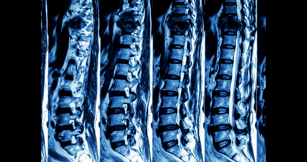
- Imaging organs and soft tissue internal structure (see the image of the spine scan to right).
- Tissue differences between normal and abnormal tissue
- Radiation imaging is not possible
What is CT scanning the Spine?
A medical diagnostic test called computed imaging (or CAT scan) is one that generates multiple images of the inside. A CT scan is painless and quick. It takes only 5-20 minutes to complete. The CT images of internal organs, bones, and soft tissue as well as those of blood vessels provide greater detail than traditional x rays. CT shows the bony structure of the spine vertebrae and intervertebral disks. You can arrange cross-sectional CT scan images in different planes or even create three-dimensional images.
CT is great for:
- Take a picture of bone, soft tissue and blood vessels simultaneously
- Pinpointing bony structures (injuries).
- Evaluation of chest and lung problems (see the image of the lung scan to right).
- Cancer detection
- Imagine patients with metal (no magnetic)
What are the differences between an MRI and a CT SCAN?
An MRI is different from a CAT scan. Also known as a CT scan, or a computed axial tomography scan, an MRI is not. It doesn’t use radiation. MRI scans can image water-containing tissues more accurately than traditional x rays. An MRI scan can identify soft tissue problems such as bulging discs and pinched nerves. MRIs can be used in cases where X-rays aren’t appropriate, such as pregnant women. People with metallic implants may not be able to undergo an MRI due to the magnetic field created by the test. A CT scan is better than an MRI for imaging calcified tissue such as bone. CT scans can also be used for diagnosing fractures and osteoarthritis.
A CT scan could reveal spinal conditions that can be diagnosed.
A spinal CT scan is a great way to diagnose neck and back pain. It shows details of bones, muscles, organs, and can even show the patient how they are feeling.
- Vertebral fractures
- Spinal degenerative changes
- Vertebral instability
- Spinal osteomyelitis
- Spinal mass and cancer
CT allows for the visualization of the entire cervical spine (neck), or the entire spine. This view is preferred by emergency departments when they need to evaluate trauma injuries in detail. CT is the most preferred imaging method for gunshot wounds due to the metal nature of the bullet. This scan provides detailed information that is not possible with other specialized scans, such as an MRI scan or magnetic resonance imaging scan (MRI). The scans may not show enough of the tendons, ligaments, and spinal cords that computed tomography can capture. These conditions are less apparent on a CT scan.
Structure and anatomy of the spine
- The spinal column is made up of 33 vertebrae. These are separated by spongy disks, and then classified into different areas.
- The cervical area is made up of seven vertebrae.
- The chest region of the thoracic region contains 12 vertebrae.
- The lower back of the lumbar area contains five vertebrae.
- The sacrum is made up of five small fused vertebrae.
- The four coccygeal vertebrae fuse to form one bone, the tailbone or the coccyx.
- The spinal cord is an important component of the central nervous system. It can be found in the vertebral canal. It runs from the base of your skull to the upper part of your lower back. The spinal cord is surrounded by the cerebrospinal fluid and spine bones. The spinal cord transmits movement and sense signals from the brain to many reflexes.
What causes a CT scan of the spine?
To evaluate your spine’s condition, a CT scan can be performed. This includes a CT scan for herniated discs, tumors, or other lesions. This scan is particularly useful in situations where other examinations, such as X Rays and physical examinations, are not conclusive. For evaluation of the effects on spine treatment such as surgery, or any other therapy, a CT scan can be taken of the spine. A CT scan may be recommended by your doctor to check the spine.
What are the possible risks of a CT scan?
Ask your doctor questions about radiation exposure and potential risks associated with the CT procedure. Keep track of radiation exposures, including any previous CT scans and other X-rays. This will enable you to notify your doctor. Radiation exposure may be connected to radiation exposure if there have been multiple X-rays taken or treatments within a short period.
If you become pregnant, or suspect that you might be pregnant, you should notify your doctor. Radiation can cause birth defects in pregnant women. If you require a CT scan of your spine, special precautions will be taken in order to minimize radiation exposure to the baby.
Nursing mothers should wait at least 24 hours before breastfeeding after receiving contrast material injections.
Allergy reactions can be caused by exposure to contrast media. If you are allergic or sensitive to medication, please inform your doctor. Research has shown that 88% of people won’t have adverse reactions to iodinated colors. It is important to tell your doctor if there have been any adverse reactions to contrast media. A seafood allergy is not considered to be a contraindication for iodine.
Patients with kidney disease or any other condition should contact their doctor. In some cases, kidney damage can be caused by contrast media. In the last decade, contrast agents and kidney disease has been discussed more extensively. Contrast exposure can cause kidney damage in patients with kidney disease. Before receiving IV contrast, patients who are taking metformin (Glucophage), an insulin medication, should notify their doctor. Metformin (Glucophage) can cause metabolic acidosis. This is a rare condition. Metformin patients will need to stop taking the drug 48 hours after surgery. Metformin users may need to have their kidney function tested before they can resume taking the drug. Your medical condition may also pose risks. Discuss your concerns with your doctor before you go ahead with the procedure.
How do I prepare for a CT scan?
You’ll receive detailed instructions when you schedule a computed Tomography Angiography appointment (CTA).
PRECAUTIONS: Your doctor will discuss with you other options.
CLOTHING: You may be asked to wear a patient dress. If you require a gown, a gown will be provided. A locker will be provided to protect your personal belongings. You must remove all piercings and leave behind jewelry and valuables.
CONTRAST MEDIA CT can be performed with or without contrast media. Contrast media improves radiologists’ ability see inside the body.
Patients who are allergic to iodine-containing contrast media should not use them. If you have kidney problems, inform the access center representative when you make an appointment. Contrast media may not be required for the scan. An alternative imaging exam may be available.
To detail side effects and risks of contrast media injections via a small tube placed in a vein called an intravenous (IV), a consent form is required. The most common CT scan is the double contrast. You will need to have contrast media before your exam. The radiologists will be able to see more of your digestive tract if you have more contrast.
ALLERGY: Please inform your access center representative if you have ever experienced an allergic reaction to contrast media when you schedule your CT scan. IV contrast will not be given to anyone who has ever experienced severe or anaphylactic reactions to contrast media. If you have ever had mild or moderate reactions in the past to contrast media, you will need medication. These plans will be discussed with you at your appointment. Discuss with your physician any known reactions to contrast media.
If your doctor orders a CT scan without contrast, you can still eat and drink. If your doctor has given you contrast, it is best to avoid eating three hours prior to your CT scan. Clear liquids are advised. You may be asked by your doctor to take any prescription medication prior to your exam.
DIABETICS: Diabetics need to eat light breakfast or lunch three hours before the scan. Depending on your oral medication results, you may have to stop taking diabetes medication 48 hours following a CT scan. You will be given detailed instructions after your CT scan.
MEDICATION: Patients are permitted to continue with their regular medication. Your doctor might recommend a different medication depending on your medical condition.
How CT Scans Work
The x-ray study of computed tomography is also known as computed tomography. A series of x-rays is rotated in different directions around a specific body part. This results in a sequence computer-generated images. These sequential images are not like traditional x-rays and show the target tissue in great detail. The x-ray data is transmitted to a computer, which then gathers it and displays it as a series of two-dimensional images on a monitor.
Intraoperative CT
Intraoperative CT scans can be used in neurosurgery to provide real-time images during and after surgery. Intraoperative CT allows for minimally invasive surgery. This eliminates the need to have extensive or open surgeries. The intraoperative CT allows for precise screw placement during spinal fusion surgery.
Preparing for a Spinal CT
A CT scan can show artifacts when you add metal jewelry, clothing, or accessories to your outfit. These artifacts may obscure images. Patients are required to remove all jewelry and wear the hospital gown before they can scan. A CT scan can be used safely by patients with pacemakers, programmable pumps, or shunts.
Spinal CT Procedure
A spinal CT scan can be done painlessly at any hospital with a CT machine. For neck and back CT scans, the same procedure is followed. The process typically follows these steps:
- The scanner table supports the patient’s body and allows them to lie down.
- After the patient is satisfied with the treatment, the technician will exit the room and take control of the CT machine in another room.
- As the scanner moves around the scan table, the scanner slides into the large circular opening on the CT machine.
- The scanner detects the x-ray beams that are emitted by the machine and absorb them into the body. The scanner receives the x-ray beams in the form of an image, which can be viewed on a PC.
For certain types of CT studies, such as CT with myelogram, contrast dyes are necessary. This dye is used for increasing contrast and CT resolution.
What happens after a CT scan?
Your procedure may have included the use of contrast media. Side effects and reactions will be closely monitored, including itching and swelling, difficulty breathing and rash. Please notify your doctor if you experience any of the following symptoms.
If you experience any pain, discomfort or redness at your IV site after your procedure, it is important to inform your doctor. This could indicate an infection. The CT scan of your spine doesn’t require any special attention. Unless you are advised otherwise by your doctor, you can resume your normal activities and diet. Your specific circumstances may require additional or alternate advice from your doctor.
Spinal CT: The risks
Tissue damage can result from CT procedures that are exposed to ionizing radiation. Computed Tomography is about half of all medical radiation. It uses between 50 and 1000 times more radiation than traditional radiographs.
A CT scan should not be done by pregnant women unless the baby is in immediate danger. CT scans should not be performed on children under 10 years of age because radiation damage can occur to their developing organs.
Many neck and back conditions can be diagnosed using CT scans. They can be used before, during, and after spinal treatment. Radiation exposure is often more beneficial than the scan’s benefits. If there is a high risk of tissue injury and radiation exposure, CT should be avoided.
